-
By:
- cierra
- No comment
tibia/fibula fracture rehabilitation protocol pdf
Tibia and fibula fractures require a structured rehabilitation approach, often detailed in a protocol, to restore optimal function and mobility.
Effective recovery involves phased progression, addressing pain, swelling, and gradually rebuilding strength and balance.
This process aims to return individuals to their desired activity levels, minimizing the risk of future injuries.
Understanding the Fracture
Fracture comprehension is crucial; the tibia, the larger weight-bearing bone, is prone to breaks from trauma, while the fibula often accompanies tibial fractures.
These injuries range in severity – stable, displaced, open, or comminuted – impacting rehabilitation timelines.
A pilon or tibial plafond fracture, an intra-articular break of the distal tibia, presents unique challenges due to joint involvement.
Understanding the fracture pattern dictates treatment, whether non-surgical (casting) or surgical (open reduction and internal fixation – ORIF), influencing the rehabilitation pathway.
The location matters; fractures of the lower tibia (S82.3) or shaft (S82.2) require tailored approaches.
Accurate diagnosis, often utilizing diagnostic radiology (73590), is paramount for effective rehabilitation planning and achieving optimal patient outcomes.
Biopsies (20245) may be performed alongside fracture repair, adding complexity.
Types of Tibia and Fibula Fractures
Tibia and fibula fractures present in diverse forms, each demanding specific rehabilitation strategies. Displaced fractures require realignment, often through open reduction and internal fixation (CPT 27792, 27752, 27759).
Bicondylar tibial plateau fractures (CPT 27536) involve both condyles, impacting knee joint stability.
Comminuted fractures exhibit multiple bone fragments, complicating healing. Open fractures, where bone penetrates the skin, carry infection risk.
Fibula fractures, frequently occurring alongside tibial fractures, can range from isolated breaks to severe disruptions.
Pilon fractures, affecting the distal tibia, are intra-articular, impacting the ankle joint.
Fracture classification – stable versus unstable – guides weight-bearing protocols.
Understanding these distinctions is vital for tailoring rehabilitation programs and optimizing functional recovery, considering the injury’s specific characteristics.

Phase 1: Immediate Post-Fracture Care (0-2 Weeks)
Initial management focuses on immobilization via casting or bracing, pain control, and minimizing swelling through elevation and early gentle exercises.
Immobilization Techniques (Casting, Bracing)
Immobilization is crucial in the initial phase, employing either casting or bracing depending on fracture stability and patient factors. Casting, typically utilizing fiberglass or plaster, provides rigid support, preventing movement and facilitating bone healing. The cast extends beyond the fracture site, often encompassing the ankle and foot to ensure complete immobilization.
Bracing offers a dynamic alternative, allowing for controlled range of motion while still providing support. Functional braces are often used for stable fractures, enabling early weight-bearing under medical supervision. The choice between casting and bracing depends on the fracture pattern, displacement, and the surgeon’s preference. Regular monitoring is essential to assess skin integrity and neurovascular status, ensuring the immobilization device remains effective and doesn’t cause complications.
Proper application and education regarding cast or brace care are vital for optimal outcomes.
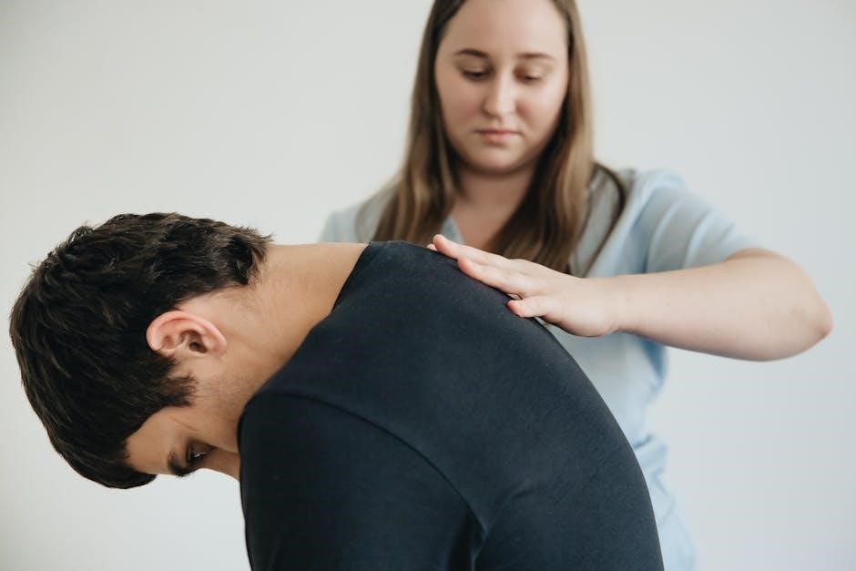
Pain Management Strategies
Effective pain management is paramount throughout tibia and fibula fracture rehabilitation. Initially, pharmacological interventions, including opioid and non-opioid analgesics, are often prescribed to control acute post-fracture pain. However, a multimodal approach is preferred, integrating medication with non-pharmacological techniques.
Elevation of the injured limb, coupled with ice application, significantly reduces swelling and pain. Range of motion exercises, initiated gently, can also alleviate discomfort by promoting circulation and preventing stiffness. As pain subsides, a transition towards over-the-counter analgesics and physical therapy modalities, such as transcutaneous electrical nerve stimulation (TENS), is encouraged.
Patient education regarding pain expectations and self-management strategies is crucial for empowering individuals to actively participate in their recovery.
Early Range of Motion Exercises (Ankle Pumps, Toe Wiggles)
Initiating early range of motion (ROM) exercises is vital, even during immobilization, to prevent stiffness and maintain circulation. Ankle pumps – repeatedly pointing toes up and down – are foundational, enhancing blood flow and reducing swelling. Similarly, toe wiggles, involving curling and extending toes, maintain neuromuscular function.
These exercises should be performed frequently throughout the day, within a pain-free range. Gentle, controlled movements are key; avoid forceful motions that could compromise the fracture site. As tolerated, incorporate alphabet tracing with the foot, promoting multi-planar movement.
The goal is to preserve joint mobility and prepare for more advanced exercises as healing progresses. Consistent, gentle ROM exercises lay the groundwork for a successful rehabilitation outcome.

Phase 2: Early Rehabilitation (2-6 Weeks)
This phase focuses on controlled weight-bearing, initiating strengthening with isometric exercises, and diligently managing swelling to promote healing and function.
Weight-Bearing Progression
Weight-bearing progression is a crucial component of early rehabilitation, carefully guided by fracture stability and pain levels. Initially, patients typically begin with non-weight-bearing or toe-touch weight-bearing, utilizing assistive devices like crutches or a walker.
As healing progresses, a gradual increase in weight-bearing is implemented. This often starts with partial weight-bearing (25-50%), progressing to weight-bearing as tolerated (WBAT), and ultimately, full weight-bearing without assistive devices.
The rate of progression varies significantly based on fracture type, individual healing capacity, and adherence to the rehabilitation protocol. Frequent monitoring by a physical therapist is essential to ensure safe and effective advancement. Pain is a key indicator; any increase in pain signals the need to reduce weight-bearing and reassess the program.
Proper biomechanics and gait training are integrated throughout this phase to prevent compensatory movement patterns and optimize functional recovery.
Strengthening Exercises (Isometric Exercises)
Isometric exercises form the foundation of early strengthening, minimizing stress on the healing fracture while activating key muscle groups. These exercises involve contracting muscles without joint movement, building strength and preventing atrophy.
Common isometric exercises include quadriceps sets (tightening the thigh muscle), hamstring sets (pressing the heel into the bed), and gluteal sets (squeezing the buttocks). Ankle isometric exercises, performed in all directions (dorsiflexion, plantarflexion, inversion, eversion), are also vital.
Focus is placed on maintaining contraction for 5-10 seconds, repeating multiple times throughout the day. Isometric exercises are generally pain-free and can be initiated early in the rehabilitation process, even while immobilized.
As tolerance improves, these exercises can be progressed by increasing hold times or adding resistance with towels or pillows. They prepare the muscles for more dynamic strengthening activities later in rehabilitation.
Swelling and Edema Control
Managing swelling (edema) is crucial throughout tibia and fibula fracture rehabilitation, directly impacting pain levels and range of motion. Initial control involves the RICE protocol: Rest, Ice, Compression, and Elevation.
Ice application, for 15-20 minutes several times a day, reduces inflammation. Compression bandages, applied snugly but not constrictively, minimize fluid accumulation. Elevation of the leg above the heart promotes venous return.
Gentle ankle pumps and toe wiggles, initiated early, encourage lymphatic drainage. As rehabilitation progresses, active range of motion exercises further assist in reducing edema.
Monitoring the circumference of the leg can track swelling reduction. Persistent or worsening edema warrants medical attention, potentially indicating complications. Effective edema control optimizes the healing environment and facilitates functional recovery.

Phase 3: Intermediate Rehabilitation (6-12 Weeks)
This phase focuses on building strength, restoring proprioception, and improving gait mechanics through progressive exercises and balance training.
Progressive Resistance Training
As pain and swelling subside during the intermediate phase (6-12 weeks), progressive resistance training becomes crucial for restoring lower extremity strength. Begin with light resistance, utilizing elastic bands or bodyweight exercises, focusing on controlled movements.
Exercises should target key muscle groups, including calf raises, hamstring curls, quadriceps sets, and hip abduction/adduction. Gradually increase resistance and repetitions as tolerated, monitoring for any signs of pain or re-injury. Ankle dorsiflexion and plantarflexion exercises with resistance bands are also beneficial.
Introduce weight-bearing exercises, such as partial squats and lunges, ensuring proper form and alignment. Prioritize functional movements that mimic daily activities. Consistent adherence to a tailored resistance training program is vital for achieving optimal strength gains and preparing for advanced rehabilitation.
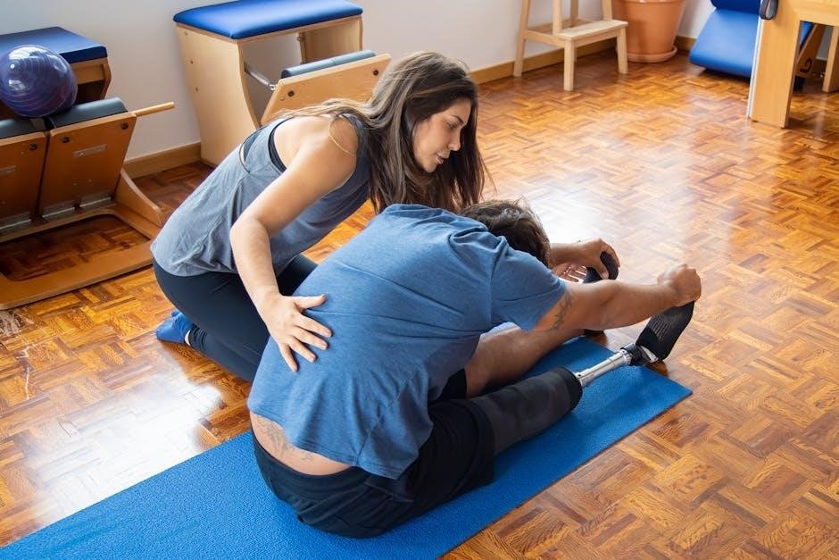
Proprioceptive Exercises
Proprioception, or the body’s awareness of its position in space, is often impaired following a tibia and fibula fracture. Re-establishing this sense is vital for regaining balance and preventing re-injury. Initiate proprioceptive training during the intermediate rehabilitation phase (6-12 weeks).
Begin with simple exercises like single-leg stance, progressing to more challenging activities such as balancing on an unstable surface (foam pad or wobble board). Incorporate dynamic movements, like reaching in different directions while maintaining balance.
Exercises should focus on improving joint position sense and neuromuscular control. Closed-chain exercises, where the foot is in contact with the ground, are particularly effective. Consistent practice enhances proprioceptive abilities, contributing to improved stability and functional performance.
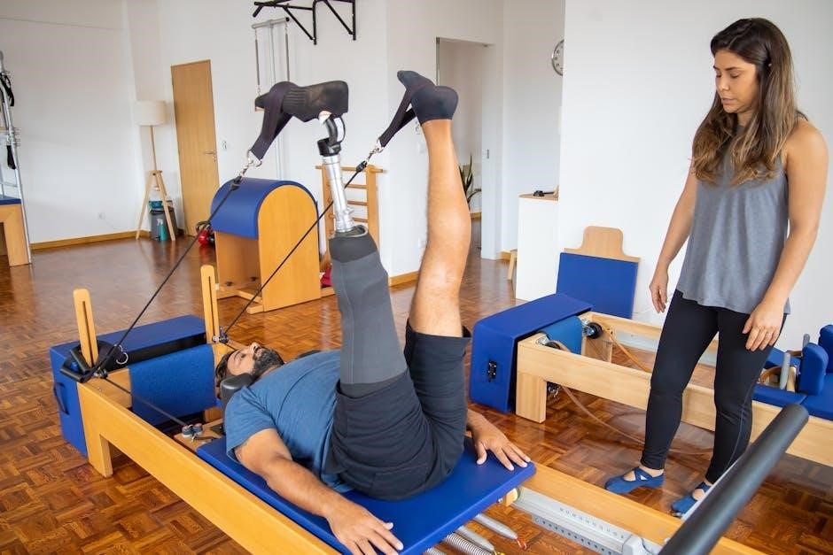
Gait Training and Balance Exercises
Restoring a normal gait pattern and dynamic balance is a crucial component of tibia and fibula fracture rehabilitation, typically addressed in the intermediate (6-12 weeks) and advanced phases. Gait training begins with assisted ambulation, utilizing assistive devices like crutches or a walker, gradually reducing reliance as strength and stability improve.
Balance exercises progress from static to dynamic activities. Start with weight shifting, then advance to tandem stance and single-leg stance. Introduce perturbations – gentle pushes – to challenge balance reactions.
Functional exercises, such as walking on uneven surfaces or navigating obstacles, simulate real-life demands. These activities enhance neuromuscular control and prepare the individual for a return to daily activities and desired recreational pursuits.
Phase 4: Advanced Rehabilitation (12+ Weeks)
This final phase focuses on sport-specific training and functional exercises like running and jumping, ensuring a safe return to activity and preventing re-injury.
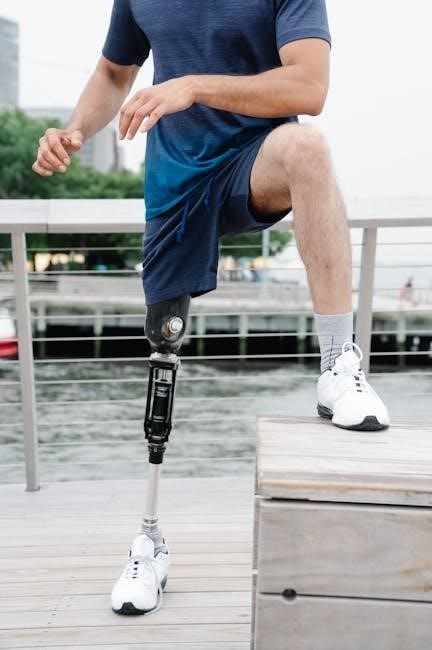
Return to Activity Specific Training
As patients progress beyond 12 weeks post-fracture, rehabilitation shifts towards replicating the demands of their specific activities. This phase necessitates a tailored approach, considering the individual’s pre-injury functional level and goals.
For athletes, this involves gradually reintroducing sport-specific drills, starting with low-impact variations and progressively increasing intensity. Running programs begin with short intervals, focusing on proper form and minimizing compensatory movements; Jumping drills are introduced cautiously, emphasizing landing mechanics and neuromuscular control.
Non-athletes benefit from exercises mimicking daily activities, such as stair climbing, walking on uneven surfaces, and lifting objects. The focus remains on restoring functional independence and confidence. Throughout this phase, continuous monitoring for pain or swelling is crucial, and adjustments are made to prevent setbacks. A successful return requires a collaborative effort between the patient, therapist, and physician.
Functional Exercises (Running, Jumping)
Advanced rehabilitation incorporates dynamic movements that challenge the tibia and fibula’s capacity to withstand impact and stress. Running drills begin with controlled intervals on even surfaces, gradually increasing speed and distance as tolerated. Emphasis is placed on proper biomechanics, including cadence, stride length, and foot strike pattern.
Jumping exercises are introduced progressively, starting with bilateral jumps and advancing to unilateral hops and jumps. Plyometric training, involving explosive movements, enhances power and agility. These exercises are carefully monitored to ensure appropriate form and minimize the risk of re-injury.
Throughout this phase, proprioceptive feedback is crucial, utilizing balance boards and agility ladders. The goal is to restore neuromuscular control and prepare the patient for the demands of their desired activities, ensuring a safe and effective return to function.
Prevention of Re-Injury
Long-term success hinges on proactive strategies to minimize the risk of re-injury following tibia and fibula fracture rehabilitation. Continued strengthening of the surrounding musculature – calves, quads, hamstrings, and core – provides dynamic stability. Maintaining flexibility through regular stretching prevents compensatory movement patterns.
Proprioceptive training remains vital, enhancing joint awareness and neuromuscular control. Gradual return to activity, avoiding sudden increases in intensity or volume, is paramount. Proper footwear and activity-specific bracing, when appropriate, offer additional support.
Patient education regarding biomechanics, injury mechanisms, and warning signs empowers self-management. Adherence to a home exercise program ensures sustained gains and promotes long-term bone health, safeguarding against future fractures.

Coding and Billing Considerations
Accurate coding is crucial for billing fracture repairs (27792, 27752, 27759) and bone biopsies (20245), alongside appropriate ICD-10 codes (S82.3, S82.2).
CPT Codes for Tibia and Fibula Fracture Repair (27792, 27752, 27759)
CPT code 27792 represents open reduction and internal fixation of a tibial plafond fracture, often a pilon fracture, involving the distal tibia’s articular surface. This procedure addresses complex intra-articular injuries requiring precise anatomical restoration.
Code 27752 covers open treatment of tibial shaft fractures, encompassing various techniques like nailing or plating to stabilize the bone. The specific approach depends on fracture characteristics and patient factors.
CPT code 27759 is utilized for open treatment of fibular shaft fractures, frequently performed in conjunction with tibial fracture repair. It ensures proper alignment and stability of the fibula, contributing to overall leg biomechanics.
Correct application of these codes is vital for accurate reimbursement, reflecting the complexity and skill involved in tibia and fibula fracture management. Documentation must clearly support the procedures performed.
ICD-10 Codes for Tibia Fractures (S82.3, S82.2)
ICD-10 code S82.3 specifically denotes a fracture of the lower end of the tibia, frequently associated with pilon fractures or injuries to the tibial plafond. This code accurately reflects fractures impacting the distal tibial articulation.
Code S82.2 identifies a fracture of the tibial shaft, encompassing breaks along the tibia’s main body. This is a common injury pattern resulting from direct trauma or rotational forces applied to the leg.
Accurate ICD-10 coding is crucial for establishing medical necessity and facilitating appropriate billing for fracture care. These codes provide essential diagnostic information for healthcare providers and payers.
Proper documentation detailing the fracture location and characteristics is paramount to ensure the correct ICD-10 code is assigned, supporting accurate claims processing and data analysis.
Bone Biopsy Coding (20245)
CPT code 20245 represents a bone biopsy procedure, specifically involving the acquisition of tissue samples for pathological examination. This code is utilized when a surgeon obtains a bone specimen, such as from the distal tibia or talus, to diagnose underlying conditions.
If multiple biopsies are performed during the same session, on different bones – for example, the tibia and the talus – each biopsy should be reported with a separate unit of code 20245.
Accurate coding requires clear documentation outlining the specific bone(s) biopsied and the clinical indication for the procedure. This ensures appropriate reimbursement and data collection.
The use of 20245 is essential when evaluating potential complications, infections, or neoplastic processes related to fractures or bone healing.

Diagnostic Imaging
Radiological assessments, utilizing CPT code 73590, are crucial for initial fracture diagnosis and monitoring healing progression throughout rehabilitation.
Role of Diagnostic Radiology (73590)

Diagnostic radiology, specifically utilizing CPT code 73590, plays a pivotal role in both the initial assessment and ongoing monitoring of tibia and fibula fractures during rehabilitation.
Initially, imaging confirms the fracture’s presence, type (e.g., pilon fracture of the tibial plafond), and severity, guiding treatment decisions. Post-reduction and internal fixation, serial radiographs are essential to evaluate alignment and stability.
Throughout rehabilitation, imaging helps track callus formation, bone healing progression, and identify any potential complications like non-union or malunion.
This allows for timely adjustments to the rehabilitation protocol, ensuring optimal outcomes. Advanced imaging modalities, while not always necessary, may be employed to assess soft tissue injuries or complex fracture patterns.
Regular imaging provides objective data to support clinical findings and optimize patient care.